Perthes Disease

He runs a biomechanical assessment clinic, advises on footwear and custom orthotics.
- Best Asics Shoes for Flat Feet - October 25, 2024
- Best Running Shoes for Flat Feet - October 22, 2024
- Posterior Tibial Tendonitis - October 21, 2024
What is Perthes Disease?
Legg-Calve-Perthes disease is named after the three doctors who discovered it. It affects young children, most commonly children aged 4 to 10 years. It is much more common in boys, five times more likely than in girls. It usually affects one hip. Both hips are affected in only 10% to 15% of all cases.
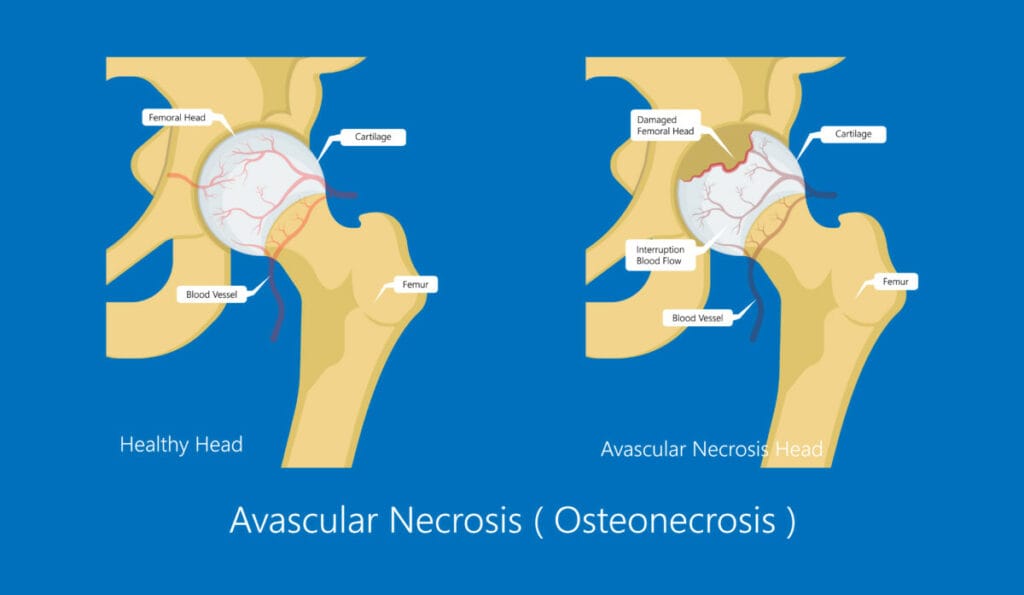
The condition results in avascular necrosis of the hip in children, leading to it becoming misshapen. When this occurs in adults, it is known as Avascular Necrosis of the hip, which can lead to a Total Hip Replacement.
Causes and Symptoms
The exact cause of Perthes disease is not known. It is thought that there may be a genetic link. More research is needed in this area.
Symptoms are initially pain that worsens with activity and improves with rest. The pain often causes a limp and restricted hip mobility. It is usually located at the hip, into the groin, sometimes into the upper thigh and occasionally as far as the knee.
Diagnosis
Perthes is diagnosed with a physical examination and confirmed with an x-ray scan.
The doctor will assess the range of movement of the hip, particularly adduction, abduction and rotation. If there is pain and restricted mobility, further confirmation will be examined with an x-ray.
Perthes Disease Stages
Necrosis
It can last several months.
There will be signs of pain, such as a limp when the child walks. Blood flow disruption and bone cells die, causing inflammation and pain
Fragmentation
Lasts 1-2 years.
The dead bone is reabsorbed and replaced with a softer bone, and the head of the femur is at risk of collapse and can flatten. This stage is called fragmentation, as the head of the femur looks like it is in pieces under X-ray imaging.
Reossification
The longest stage can last several years.
The soft bone initially formed to replace the necrosed tissue is gradually replaced with harder bone tissue. This all occurs under the articular cartilage.
Healed
Once the re-ossification has been completed, the femoral head will be healed and remain in the shape it has now formed. Ideally, the head of the femur will be round to fit in the socket. This will depend on the extent of the damage and age of onset of Perthes. Older children will have less potential for bone regrowth and will likely have poorer outcomes.
Treatment
Monitoring the progression of Perthese disease is essential for appropriate treatment. Repeated X-ray images are needed regularly throughout the disease process and recovery. In mild cases with minimal changes to the femoral head, minimal treatment is necessary, but the hip must be monitored, and pain relief provided if necessary.
Pain relieving medication is usually given in the form of non-steroidal anti-inflammatories. The death and breakdown of the tissue cause inflammation of the hip. Therefore, this is the most effective pain relief.
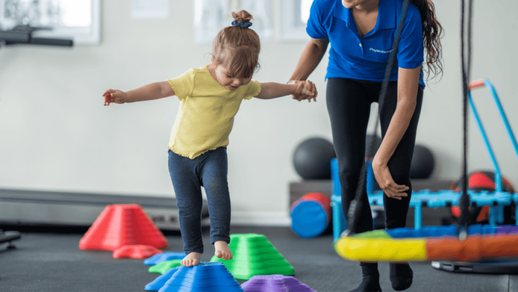
Non-surgical treatment focuses on activity modification and physical therapy to maintain hip mobility. Restricting activity mainly reduces impact and weight through the hip to protect the shape of the femoral head. In moderate to severe cases, crutches or a walker will be needed to reduce weight bearing.
Casting and Bracing for Perthes Disease
If there is evidence of extensive damage and deformity of the femoral head developing, your doctor will likely recommend a brace or cast. Petrie braces or casts keep the legs straight and spread wide in an abducted position.
In this position, the femoral head is in the best position within the joint socket to heal well and in good shape. The caste will usually be worn for 4-6 weeks and may need to be repeated.
Surgery
Sometimes, the adductor longus muscle along the inside of the thigh is too tight. It restricts the mobility of the hip and may need to be released with a minor surgical procedure called a tenotomy. This is sometimes needed before the Petrie cast can be applied.
Surgical treatments are more commonly needed if the femoral head has significant damage to more than 50% of the bone or if non-surgical treatments have been unsuccessful.
An osteotomy is a standard surgical treatment for Perthes disease. This procedure uses pins and a plate to hold the femoral head in position to allow successful healing.
These are removed once healing is complete. The prognosis is worse for children over 8 years old, and they will more likely need surgical treatment.
Recovery and Outcomes of Perthes Disease
In most cases, the child will recover well and have no further issues into adulthood. However, if the case was severe or the child developed symptoms at an older age, after 8 years, there is a worse outcome, and issues are more likely to continue into adulthood. The most common issue is the early onset of hip osteoarthritis.
_______________________________________________________________________
We are specialists in treating knee conditions such as a Perthes Disease, and you can see one of our Paediatric Physiotherapists in our clinic in Fulham, South West London.
We offer Online Appointments for £60 and Face-to-Face appointments for £85 in our clinics.
Related Articles
How Long After Hip Replacement Before Skiing – Hip Pain Diagram – Hip Pain Running